
诊断 / 测量研究Diagnostic / measurementJAMA Oncology · 2020 · 6(10): 1581–1588
External Evaluation of 3 Commercial Artificial Intelligence Algorithms for Independent Assessment of Screening Mammograms
三种商用人工智能算法独立判读筛查乳腺 X 线片的外部评估
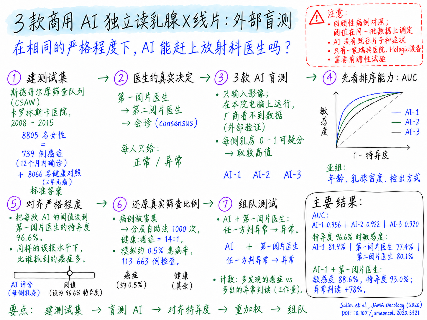
斯德哥尔摩一支独立团队在 8805 例筛查检查(739 例癌症、8066 名健康对照)上盲法运行了 3 款商用乳腺 X 线 AI 算法。表现最好的 AI-1 的 AUC 为 0.956;在与第一阅片医生相同的 96.6% 特异度下,其敏感度为 81.9%,第一阅片医生为 77.4%。AI-1 与第一阅片医生组合检出的癌症最多(敏感度 88.6%),但特异度降至 93.0%。
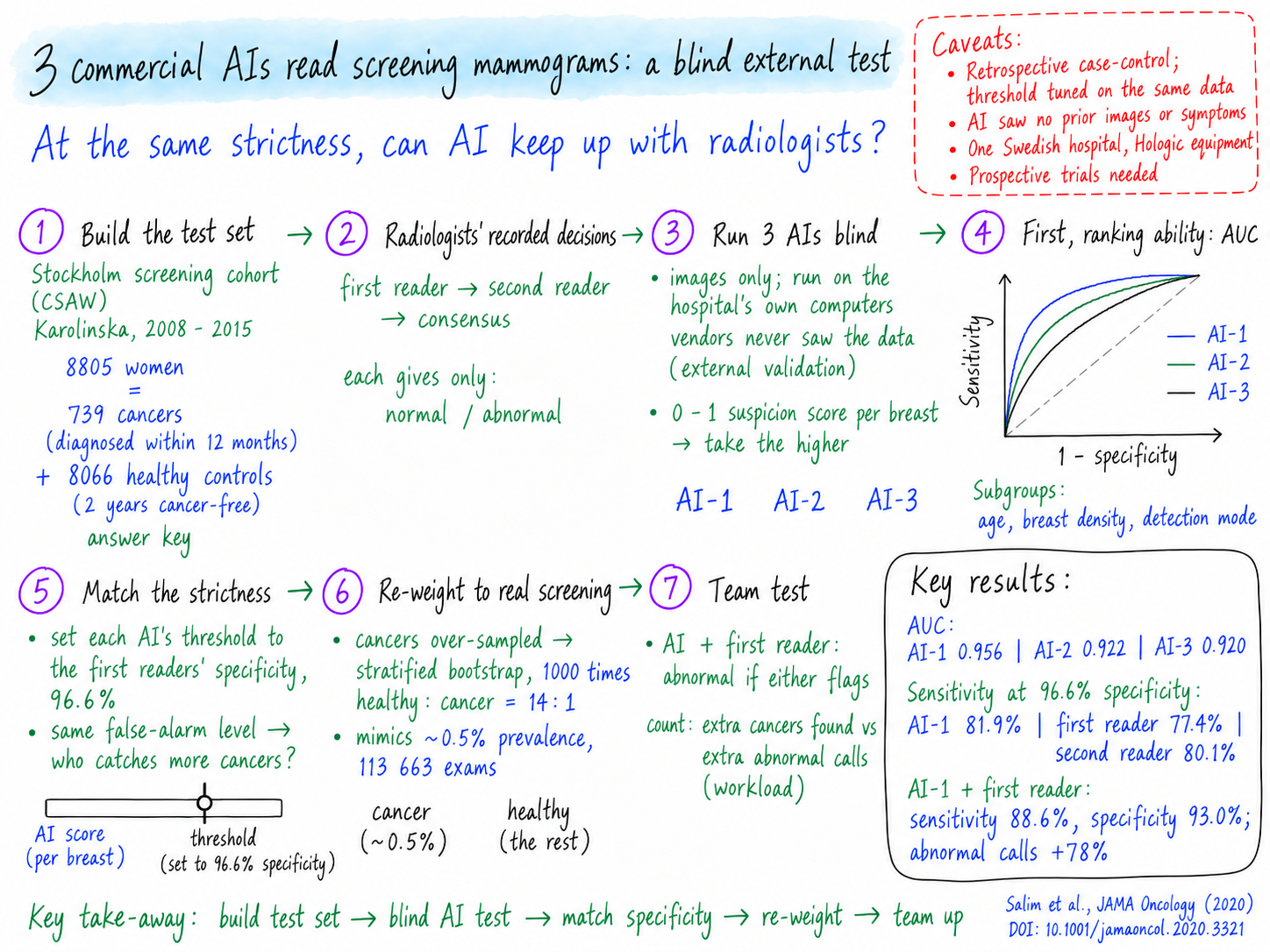
An independent team in Stockholm ran 3 commercial mammography AI algorithms blind on 8805 screening exams (739 cancers, 8066 healthy controls). The best one (AI-1) reached an AUC of 0.956 and, at the first readers' specificity of 96.6%, a sensitivity of 81.9% versus 77.4% for first-reader radiologists; pairing AI-1 with the first reader found the most cancers (88.6% sensitivity) but at a lower specificity of 93.0%.
方法图解Visual method notes
研究背景Background
- 问题Problem
- 人群乳腺 X 线筛查使乳腺癌死亡率下降了 20%–40%,但放射科医生工作量大,不同阅片者的判读质量差异明显。Population mammography screening has cut breast cancer mortality by 20% to 40%, but radiologist workload is high and reading quality varies between readers.
- 研究空白Gap
- 已有深度学习计算机辅助检测(CAD)研究报告了接近放射科医生的水平,但常常使用非筛查人群、对照医生表现偏弱,或算法并未上市;而且没有一项研究由第三方对多款相互竞争的商用算法做外部验证和比较。Deep-learning CAD studies reported radiologist-level performance, but they often used non-screening populations, weak human comparators, or algorithms not on the market — and none had a third party externally validate and compare competing commercial algorithms.
- 研究目标Objective
- 对 3 款已商用的 AI CAD 算法作为独立阅片者进行外部评估,并评估它们与放射科医生组合时的筛查表现。To externally evaluate 3 commercially available AI CAD algorithms as independent mammography readers, and to assess screening performance when they are combined with radiologists.
 ELLE · 阅读向导 / READING COMPANION
把概念连起来,再往下读 / Take a moment to connect the ideas
和 Elle 一起理清基础概念 / A few foundations, with Elle
需要时展开术语解释,再用一个小问题检验理解。 / Open a term when you need it, then try one quick question.
敏感度与特异度 / Sensitivity and specificity
敏感度:有癌症的女性中被判为异常的比例。特异度:健康女性中被正确判为正常的比例。提高一个,通常会降低另一个。 / Sensitivity: the share of women with cancer that a reader flags. Specificity: the share of healthy women correctly called normal. Raising one usually lowers the other.
AUC / AUC
在所有可能的阈值下,评分把有癌症的女性排在无癌女性之前的能力。它不是判断正确的百分比。 / How well a score ranks women with cancer above women without, across every possible threshold. It is not the percentage of correct calls.
外部验证 / External validation
由独立于开发者的团队,用模型从未见过的地方的数据来检验一个已完成的模型。 / Testing a finished model on data from a place it never saw, by a team independent of the developers.
研究方法Methods
研究目标是:由与产品无利益关系的第三方判断,现在市面上的 AI 程序能否像放射科医生一样判读筛查乳腺 X 线片。研究者从斯德哥尔摩筛查队列中构建测试集:739 名在筛查后 12 个月内确诊癌症的女性,以及 8066 名随机抽取、随访 2 年无癌的女性。他们在自己的电脑上运行三款匿名商用 AI 程序,厂商从未接触数据;再把每个程序 0–1 的可疑度评分,按照与一读医生同样的严格程度(特异度 96.6%)转换为正常/异常判断。随后比较每个 AI 和每位医生分别抓到多少癌症,把样本重新加权成接近真实筛查人群的病例构成,并模拟 AI 与医生搭档的各种组合,看哪种组合能以最少的异常判读找到最多的癌症。
The goal was to find out whether any AI program sold today can read screening mammograms as well as a radiologist, judged by someone with no stake in the products. The researchers built a test set from a Stockholm screening cohort: 739 women whose cancer was confirmed within 12 months of a screening exam and 8066 randomly chosen women who stayed cancer-free for 2 years. They ran three anonymous commercial AI programs on the images on their own computers, so the companies never saw the data, and turned each program's 0–1 suspicion score into a normal/abnormal call by matching the strictness of the first-reader radiologists (96.6% specificity). They then compared how many cancers each AI and each radiologist caught, re-weighted the sample toward a real screening case mix, and simulated teams of AI plus radiologists to see which combination found the most cancers for the fewest abnormal calls.
研究速览Study at a glance
研究对象Population
2008–2015 年在卡罗林斯卡接受筛查的 40–74 岁女性 8805 人,无假体、无乳腺癌史:癌症病例 739 人,随机健康对照 8066 人。
8805 women aged 40–74 screened at Karolinska 2008–2015, no implants or prior breast cancer: 739 cancer cases, 8066 random healthy controls.
数据来源Data source
CSAW 队列(Hologic 数字乳腺 X 线图像),关联区域筛查登记库和乳腺癌质量登记库。
CSAW cohort (Hologic digital mammograms) linked to regional screening and breast cancer quality registers.
干预 / 暴露Intervention / exposure
待评价检测:3 款匿名商用 AI CAD 算法,仅读图像,每侧乳房评分 0–1;检查评分取两侧较高者。
Index tests: 3 anonymous commercial AI CAD algorithms reading images only, score 0–1 per breast; exam score = max of both breasts.
对照Comparator
双人阅片项目中记录在案的一读、二读和会诊决定。
Recorded first-reader, second-reader and consensus decisions of radiologists in the double-reading program.
结局指标Outcomes
筛查时或 12 个月内病理确诊癌症(阴性:2 年无癌);AUC、敏感度、特异度、检出率与异常判读率。
Pathology-confirmed cancer at screening or within 12 months (negative: 2-year cancer-free); AUC, sensitivity, specificity, detection and abnormal-interpretation rates.
分析方法Analysis
AUC 与 DeLong 检验;切点匹配一读特异度 96.6%;分层 bootstrap(1000 次,14:1);AI–医生组合规则;Stata 15.1。
AUC with DeLong tests; cut-off matched to first-reader specificity 96.6%; stratified bootstrap (1000, 14:1); AI–reader combination rules; Stata 15.1.
场景与时间Setting & period
瑞典斯德哥尔摩卡罗林斯卡大学医院;2008–2015 年人群筛查。
Karolinska University Hospital, Stockholm, Sweden; population-based screening 2008–2015.
方法分步讲解Step by step
- 1
构建测试集 · Build the test set
输入:斯德哥尔摩筛查队列(CSAW)。团队纳入筛查后 12 个月内确诊乳腺癌的全部女性(“病例”),再随机抽取随访 2 年无癌的健康女性(“对照”),最终 8805 人:739 例病例、8066 名对照。这就像出一份带标准答案的考卷:每位女性是否真有癌,由病理和登记库随访确认,而不是由任何阅片者决定。与真实筛查日不同,这里癌症被刻意“放大”(病例对照抽样,C5),所以后面几步必须校正。Input: a Stockholm screening cohort (CSAW). The team took every woman diagnosed with breast cancer within 12 months after a screening exam (the 'cases'), plus a random draw of healthy women who stayed cancer-free for 2 years (the 'controls'), ending with 8805 women: 739 cases and 8066 controls. It is like building an exam paper with a known answer key: whether each woman truly had cancer was confirmed by pathology and registry follow-up, not by any reader. Unlike a real screening day, cancers are deliberately over-represented (case-control sampling, C5), so later steps must correct for that.为什么这样做Why this step在给任何人打分之前,先要有一份独立于 AI 和医生的可靠标准答案。A reliable answer key, independent of both the AI and the radiologists, is needed before anyone can be graded.技术细节Technical detail · M1, M3, M6, M7, M9, M10, M11, M15来自 CSAW(卡罗林斯卡,2008–2015,Hologic 全视野数字乳腺摄影)的回顾性病例对照样本:病例 = 筛查时或 12 个月内病理确诊(排除 >12 个月的 419 次检查);对照 = 随机 10 000 名健康女性经排除(要求 2 年无癌随访);最终 N = 8805(739/8066)。Retrospective case-control sample from CSAW (Karolinska, 2008–2015, Hologic FFDM): cases = pathology-confirmed cancer at screening or ≤12 months (419 exams >12 months excluded); controls = random 10 000 healthy women with exclusions (2-year cancer-free follow-up required); final N = 8805 (739/8066). - 2
调出医生的真实判读 · Retrieve radiologists' real decisions
输入:筛查登记库。团队调出每份检查当年医生的真实判断:一读、二读(通常更有经验,且能看到一读结果)以及会诊决定。每个判断都是简单的“正常/异常”,所以可以用敏感度和特异度(C1)来打分。可以把它想成直接使用医生“当年已交的答卷”,而不是请他们重新考一次。Input: the screening register. For each exam the team pulled what the radiologists actually decided at the time, years earlier: first reader, second reader (often more experienced and able to see the first reader's call), and the consensus meeting. Each decision is a simple normal/abnormal, so it can be scored with sensitivity and specificity (C1). Think of it as using the human doctors' already-submitted exam answers rather than asking them to retake the test.为什么这样做Why this step真实临床决策提供了贴近现实的人类基准,而不是实验室阅片研究。Real-world clinical decisions give a realistic human benchmark instead of a lab reader study.技术细节Technical detail · M12, M13, M14从斯德哥尔摩-哥特兰区域癌症中心登记库提取 25 位一读、20 位二读及会诊(正常/召回)的前瞻性二分类判读,通过个人身份号码关联。Prospectively recorded binary assessments of 25 first readers, 20 second readers and consensus (normal/recall) from the Regional Cancer Center Stockholm-Gotland register, linked via personal identity numbers. - 3
三款商用 AI 盲测 · Run three commercial AIs blind
输入:只有乳腺 X 线图像。三家厂商的算法(AI-1、AI-2、AI-3)在研究者自己的电脑上运行;厂商从未看到图像,也不参与分析——这就是外部验证(C4)。每个 AI 给每侧乳房打 0–1 的可疑度分,两侧中较高的分作为这位女性的分数。就像三名外校学生参加同一场封闭考试,考场里没有他们学校的人。Input: the mammogram images only. The three vendor algorithms (AI-1, AI-2, AI-3) were run on the researchers' own computers; the companies never saw the images or took part in the analysis — this is external validation (C4). Each AI gives every breast a suspicion score from 0 to 1, and the higher of the two breasts becomes the woman's score. It is like three outside students sitting the same sealed exam, with nobody from their school in the room.为什么这样做Why this step防止厂商针对测试数据做调整,使评分反映产品面对新病人时的真实表现。Keeps vendors from tuning to the test data, so the scores reflect how the products would really perform on new patients.技术细节Technical detail · M2, M16, M17, M183 款匿名商用深度学习 CAD 产品(未获 FDA 独立阅片批准)在本地仅用图像推理;每侧乳房评分 0–1;检查层面评分 = max(左, 右)。Images-only inference, locally, by 3 anonymous commercial deep-learning CAD products (not FDA-approved as independent readers); per-breast score 0–1; exam-level score = max(left, right). - 4
用 AUC 检查排序能力 · Rank-quality check with AUC
输入:每个 AI 的评分加上标准答案。在选任何切点之前,团队先看每个 AI 把患癌女性排在未患癌女性前面的能力,用 AUC(C7)概括,包括总体和亚组(年龄、乳腺密度、癌症是在筛查时发现还是之后在临床上发现)。DeLong 检验(C9)判断一个 AI 的 AUC 是否真的高于另一个。就像先比较三位评委给选手排名次的水平,再决定及格线放在哪里。Input: each AI's scores plus the answer key. Before picking any cut-off, the team asked how well each AI ranks women with cancer above women without, summarized as the AUC (C7), both overall and in subgroups (age, breast density, and whether the cancer was found at screening or later as a clinically detected cancer). The DeLong test (C9) checks whether one AI's AUC is really higher than another's. It is like comparing how well three judges order contestants, before deciding where the pass mark goes.为什么这样做Why this stepAUC 不依赖某一个阈值,可以公平地比较各个 AI。AUC compares the AIs fairly without depending on any single threshold.技术细节Technical detail · M8, M19, M20, M25每个算法计算总体 AUC,以及按年龄、乳腺密度(4 个体位均值,以中位数划分)和检出方式分组的 AUC;两两 AUC 差异用 DeLong 检验;置信区间用三明治方差估计量;次要分析加入筛查后 12–23 个月确诊的 174 例癌症。AUC per algorithm overall and by age, mammographic density (4-view mean, split at median) and detection mode; pairwise AUC differences by DeLong test; CIs by sandwich variance estimator; secondary analysis adding 174 cancers diagnosed 12–23 months after screening. - 5
让 AI 与医生同样严格 · Match AI to radiologists' strictness
输入:AI 的连续评分和一读医生的特异度。医生给出“是/否”,而 AI 给出一个数字,所以团队为每个 AI 选了一个切点(C6),让它与一读医生一样“严格”——把约 96.6% 的健康女性判为正常。然后才能问一个公平的问题:在误报水平相同的情况下,谁抓到的癌更多?就像先把两个烟雾报警器的误报率调成一样,再看哪个能发现更多真实火情。Input: continuous AI scores and the first readers' specificity. Doctors give yes/no, but the AI gives a number, so the team picked a cut-off (C6) for each AI that makes it as 'strict' as the first-reader radiologists — calling about 96.6% of healthy women normal. Then they could ask the fair question: at the same false-alarm level, who catches more cancers? It is like setting two smoke detectors to the same false-alarm rate before seeing which one detects more real fires.为什么这样做Why this step如果没有统一的工作点,更高的敏感度可能只是因为误报更多。Without a common operating point, a higher sensitivity could simply mean more false alarms.技术细节Technical detail · M21, M23每个评分在特异度最接近一读 96.6% 的切点处二分类;指标:敏感度、特异度、准确率、异常判读率、癌症检出率、假阴性率(每 1000 人)和 PPV,对每个 AI 及一读、二读和会诊分别计算。Each score dichotomized at the cut point giving specificity closest to first readers' 96.6%; metrics: sensitivity, specificity, accuracy, abnormal interpretation rate, cancer detection rate, false-negative rate (per 1000) and PPV, for each AI and for first reader, second reader and consensus. - 6
按真实患病率重新加权 · Re-weight to real screening prevalence
输入:富集后的样本。因为癌症被放大了(S1),像“每 1000 名筛查女性检出几例癌”这样的数字会失真。团队用分层自助重抽样(C8):1000 次分别按 14:1 的比例重新抽取健康女性和癌症病例,目标是模拟真实筛查人群(约 0.5% 为筛查检出癌)。就像把样本重新调配,让比例重新接近真实人群——但它只能重复使用研究中已有的女性,不能加入新类型的人。Input: the enriched sample. Because cancers were over-represented (S1), numbers like 'cancers found per 1000 women screened' would be misleading. The team used stratified bootstrap resampling (C8): 1000 times they redrew healthy women and cancer cases separately at a 14:1 ratio, aiming to mimic the real screening population (about 0.5% screen-detected cancer). It is like re-mixing a sample so the proportions look like the real crowd again — but it can only reuse the women already in the study, not add new kinds of women.为什么这样做Why this step依赖患病率的指标(每 1000 人比率、PPV)只有在真实的病例构成下才有意义。Prevalence-dependent metrics (rates per 1000, PPV) are only meaningful at a realistic case mix.技术细节Technical detail · M22分层 bootstrap,1000 次抽样,健康:癌症 = 14:1,以近似原始队列(约 0.5% 为筛查检出癌)。Stratified bootstrapping, 1000 samples, healthy:cancer = 14:1, to approximate the source cohort (~0.5% screen-detected cancer). - 7
测试 AI 与医生搭档 · Test AI–radiologist teamwork
输入:各 AI 和医生的“是/否”判断。团队模拟了新的阅片组合:每个 AI 与一读、二读或两位医生搭配(任一方标记即为异常);三个 AI 联合(任一 AI 标记即异常);全部 AI 加医生(至少 2 方标记才算异常)。对每种组合统计检出的癌症数与异常判读数(召回工作量)。就像测试把两人小组中的一个人换成机器,能否发现更多问题、又要付出多少误报代价。Input: the yes/no calls from the AIs and the readers. The team simulated new reading teams: each AI paired with the first reader, second reader or both (abnormal if anyone flags it); all three AIs together (abnormal if any AI flags); and all AIs plus radiologists (abnormal if at least 2 flag). For each team they counted cancers found against abnormal calls (recall workload). It is like testing whether swapping one human member of a two-person team for a machine catches more problems, and at what cost in false alarms.为什么这样做Why this step现实中 AI 最可能替代或辅助其中一位阅片者,所以关键问题是组合后的表现,而不只是 AI 单独的表现。In practice AI would most likely replace or support one reader, so the useful question is how combined teams perform, not just AI alone.技术细节Technical detail · M24用“或”规则(≥1 方异常)模拟 AI + 一读/二读/双读以及 3 个 AI 联合,用“N 中至少 2 方”规则模拟全部 AI 加医生;比较异常判读数与检出癌数。Simulated combinations using OR-rules (≥1 abnormal) for AI + first/second/both readers and for all 3 AIs, and a ≥2-of-N rule for all AIs plus radiologists; compared abnormal interpretations vs cancers detected.
读懂方法需要的前置知识Concepts you need first
C1
敏感度与特异度(sensitivity / specificity) · Sensitivity and specificity
敏感度是真正患病的人中被检查标出来的比例;特异度是健康人中被正确判为正常的比例。通常提高一个,另一个就会下降。
Sensitivity is the share of people who truly have the disease that a test flags; specificity is the share of healthy people the test correctly calls normal. Raising one usually lowers the other.
经典应用Classic uses
- 以 PCR 为标准评价新冠抗原快速检测Rating COVID-19 rapid antigen tests against PCR
- 评价 D-二聚体检测排除血栓的能力Evaluating the D-dimer blood test for ruling out blood clots
C2
乳腺 X 线筛查与双人阅片(double reading) · Screening mammography with double reading
健康女性每 1–2 年拍一次乳房 X 光;在瑞典和欧洲很多地方,每份检查由两位放射科医生分别阅读,任何一人标记异常就进入会诊,决定是否召回做进一步检查。
Healthy women get a breast X-ray every 1–2 years; in Sweden and much of Europe two radiologists read each exam independently, and any flag leads to a group discussion that decides whether to recall the woman for more tests.
经典应用Classic uses
- 欧洲各国乳腺筛查项目(如英国 NHS 乳腺筛查计划)European national breast screening programs (e.g., the UK NHS Breast Screening Programme)
C3
深度学习计算机辅助检测(AI CAD) · Deep-learning computer-aided detection (AI CAD)
一种从大量带标签图像中学会识别模式的程序,对新图像输出“看起来有多可疑”;在本研究中它是从公司买来的“黑箱”。
A computer program that has learned from many labeled images to recognize patterns, and outputs how suspicious a new image looks; here it is a 'black box' bought from a company.
经典应用Classic uses
- 用眼底照片筛查糖尿病视网膜病变(如获 FDA 授权的 IDx-DR)Diabetic retinopathy screening from retinal photos (e.g., the FDA-authorized IDx-DR system)
- 根据照片对皮肤病变分类Skin-lesion classification from photos
用于Used in: S3
C4
外部验证(external validation) · External validation
由独立于开发者的人,用模型开发时从未见过的地方和人群的数据来测试已完成的模型,看它在真实世界里是否依然好用。
Testing a finished model on data from a place and people it never saw during development, by someone independent of the developers, to see whether it still works in the real world.
经典应用Classic uses
- 在美国以外的人群中重新检验 Framingham 心血管风险评分Re-testing the Framingham heart-risk score in non-US populations
- 密歇根大学医院对 Epic 脓毒症预警模型的独立评估Independent evaluation of the Epic Sepsis Model at Michigan Medicine
C5
病例对照(富集)抽样与患病率 · Case-control (enriched) sampling and prevalence
研究不是纳入所有筛查女性(其中只有约 0.5% 患癌),而是纳入全部癌症病例加上随机抽取的一部分健康女性,所以癌症“被放大”了;凡是依赖患病率的指标都必须再校正回来。
Instead of taking every screened woman (where only about 0.5% have cancer), the study takes all cancer cases plus a random subset of healthy women, so cancers are over-represented; numbers that depend on how common the disease is must then be corrected.
经典应用Classic uses
- Doll 和 Hill 关于吸烟与肺癌的经典病例对照研究Classic case-control studies linking smoking to lung cancer (Doll and Hill)
C6
评分阈值(工作点,operating point) · Score threshold (operating point)
连续评分要变成“是/否”,就必须选一个切点;切点放在哪里,决定了多抓癌症与多出误报之间的取舍。
A continuous score must be turned into yes/no by choosing a cut-off; where you put it decides the trade-off between catching more cancers and raising more false alarms.
经典应用Classic uses
- 选择前列腺癌检查的 PSA 切点(如 4 ng/mL)Choosing the PSA cut-off (e.g., 4 ng/mL) for prostate cancer workup
- 设定床旁监护仪的血氧报警阈值Setting SpO2 alarm limits on bedside monitors
C7
ROC 曲线与曲线下面积(AUC) · ROC curve and AUC
ROC 曲线把每个可能切点下的敏感度与误报率画出来;曲线下面积(AUC)概括了评分把病人排在健康人前面的能力,0.5 相当于抛硬币,1 为完美。
The ROC curve plots sensitivity against false alarms for every possible cut-off; the area under it (AUC) summarizes how well the score ranks sick people above healthy people, from 0.5 (coin flip) to 1 (perfect).
经典应用Classic uses
- 比较心肌梗死的诊断标志物,如肌钙蛋白Comparing diagnostic biomarkers such as troponin for heart attack
- 报告临床风险评分和机器学习模型的表现Reporting performance of clinical risk scores and ML models
用于Used in: S4
C8
自助重抽样(bootstrap) · Bootstrap resampling
从已有数据中有放回地反复抽样,每次重新计算结果,看结果波动多大;分层 bootstrap 在每个组内分别抽样,因此可以固定各组人数比例。
Repeatedly drawing new samples with replacement from the data you have, recomputing the result each time, to see how much it varies; stratified bootstrap draws from each group separately so you can fix the group sizes.
经典应用Classic uses
- 为中位数或机器学习指标计算置信区间Confidence intervals for medians or ML performance metrics
用于Used in: S6
C9
DeLong 检验 · DeLong test
一种统计检验,判断在同一批人上计算的两个 AUC 的差别是否超出偶然;它考虑了两者用的是同一批病例这一点。
A statistical test for whether two AUCs measured on the same people differ by more than chance, accounting for the fact that both were computed on the same cases.
经典应用Classic uses
- 在同一队列上比较两种诊断检测或风险模型(R 的 pROC 包)Comparing two diagnostic tests or risk models on one cohort (pROC package in R)
用于Used in: S4
方法细节(原文提取)Method details (from the paper) · 27
| M1 | 研究设计Study design | 回顾性病例对照研究:由第三方对 3 款商用 AI 计算机辅助检测(CAD)算法作为独立阅片者进行外部评估,并评估它们与放射科医生联合时的筛查表现。 Retrospective case-control study: an external (third-party) evaluation of 3 commercial AI computer-aided detection (CAD) algorithms used as independent readers of screening mammograms, and of their combination with radiologists. | p. 1–2 |
| M2 | 验证Validation | 算法开发方无法接触研究图像,也未参与评估过程。 The algorithm developers had no access to the study images and no involvement in the evaluation process. | p. 2 |
| M3 | 场景Setting | 样本来自 CSAW(瑞典筛查年龄女性队列)数据集:2008–2015 年斯德哥尔摩郡所有受邀参加筛查的 40–74 岁女性;所有检查均来自同一机构——卡罗林斯卡大学医院。 Sample drawn from the CSAW (Cohort of Screen-Aged Women) data set: all women aged 40 to 74 years in Stockholm county invited for screening between 2008 and 2015; all examinations were from one institution, Karolinska University Hospital. | p. 2 |
| M4 | 场景Setting | 斯德哥尔摩郡筛查间隔为 24 个月;2012 年以前,40–49 岁女性的间隔为 18 个月。 Screening interval in Stockholm county is 24 months; until 2012 it was 18 months for women aged 40 to 49 years. | p. 2 |
| M5 | 伦理Ethics | 经斯德哥尔摩伦理审查委员会批准,并豁免个人知情同意。 Approved by the Stockholm ethical review board, which waived individual informed consent. | p. 2 |
| M6 | 研究对象Participants | 病例组:CSAW 中 2008–2015 年确诊乳腺癌、确诊前有完整筛查检查、既往无乳腺癌且无假体植入的全部 40–74 岁女性。 Cases: all women aged 40 to 74 years in CSAW diagnosed with breast cancer 2008–2015 who had a complete screening examination before diagnosis, no prior breast cancer and no implants. | p. 2 |
| M7 | 研究对象Participants | 排除 419 次检查,因其检查日期与癌症确诊间隔超过 12 个月(筛查时癌已存在的可能性较低)。 419 examinations were excluded because more than 12 months elapsed between the examination and the cancer diagnosis (lower likelihood that cancer was present at screening). | p. 2 |
| M8 | 敏感性分析Sensitivity | 次要分析:把筛查后 12–23 个月确诊癌症的 174 名女性加入病例组。 Secondary analysis: 174 women diagnosed with cancer 12 to 23 months after screening were added to the case group. | p. 2 |
| M9 | 研究对象Participants | 对照组:随机抽取 10 000 名健康女性(为了计算效率,同时保持代表性);排除无癌随访不足 2 年者 995 人、检查晚于 2015 年 12 月 31 日者 909 人、有假体者 26 人,以及放射科医生编号未知的检查 99 次(eFigure 1)。 Controls: a random sample of 10 000 healthy women (random sampling for efficient computing while keeping representativeness); excluded 995 with <2-year cancer-free follow-up, 909 with examinations after December 31, 2015, 26 with implants, and 99 examinations with unknown radiologist ID (eFigure 1). | p. 2 |
| M10 | 样本量Sample size | 最终样本:8805 名女性——739 名确诊乳腺癌(阳性),8066 名随机抽取的健康对照(阴性)。 Final study sample: 8805 women — 739 diagnosed with breast cancer (positive) and a random sample of 8066 healthy controls (negative). | p. 1 |
| M11 | 数据Data | 所有图像均由 Hologic 全视野数字乳腺 X 线摄影设备采集;筛查要求每侧乳房拍 2 个体位。 All images were acquired on full-field digital mammography Hologic equipment; screening requires 2 views of each breast. | p. 2 |
| M12 | 对照Comparator | 对照:从斯德哥尔摩-哥特兰区域癌症中心筛查登记库中提取的、前瞻性记录的放射科医生判读。采用双人阅片,每位阅片者做“正常/异常(标记讨论)”二分类判断;任何异常判断进入会诊讨论,再做“正常/召回”二分类决定。 Comparator: prospectively recorded radiologist assessments from the Regional Cancer Center Stockholm-Gotland screening register. Double reading, each reader making a binary normal/abnormal (flagged) decision; any abnormal assessment goes to a consensus discussion with a binary normal/recall decision. | p. 2 |
| M13 | 对照Comparator | 共有 25 位一读和 20 位二读放射科医生;一读/二读没有固定分工,但二读通常更有经验,且能看到一读的判断。 There were 25 first-reader and 20 second-reader radiologists; roles were not formally designated, but the second reader was often more experienced and could see the first reader's assessment. | p. 2 |
| M14 | 数据Data | 癌症诊断、肿瘤特征和影像判读结果通过瑞典个人身份号码与区域乳腺癌质量登记库和筛查登记库关联获得。 Cancer diagnoses, tumor characteristics and radiologic assessments were obtained by linkage with the regional breast cancer quality register and screening register using Swedish personal identity numbers. | p. 2 |
| M15 | 结局Outcome | 参考标准:阳性 = 筛查时或其后 12 个月内经病理确诊的乳腺癌;阴性 = 2 年无癌随访。 Reference standard: positive = pathology-confirmed breast cancer at screening or within 12 months thereafter; negative = 2-year cancer-free follow-up. | p. 1–2 |
| M16 | 模型Model | 3 款来自不同厂商的商用 AI CAD 算法(AI-1、AI-2、AI-3)在作者本地硬件上处理图像;它们均未获 FDA 批准用作独立阅片者;厂商匿名;各厂商按作者设计的结构描述其算法(eAppendix)。 Images were processed locally on the authors' hardware by 3 commercial AI CAD algorithms (AI-1, AI-2, AI-3) from different vendors; none were FDA-approved as independent readers; vendors remained anonymous; each vendor described its algorithm using a structure devised by the authors (eAppendix). | p. 2–3 |
| M17 | 模型Model | 算法只使用图像(不用其他数据),对每侧乳房输出 0–1 的癌症可疑度评分(1 = 最可疑)。 The algorithms used the images only (no other data) and output a prediction score between 0 and 1 for suspicion of cancer for each breast (1 = highest suspicion). | p. 3 |
| M18 | 测量Measurement | 所有分析在检查层面(等同于患者层面)进行,取左右乳评分中较高者。 All analyses were at examination level (equivalent to patient level), using the maximum of the left- and right-breast scores. | p. 3 |
| M19 | 统计Statistics | 计算每个 AI 系统的总体 AUC,并按年龄、乳腺密度和检出方式(筛查检出 vs 临床检出)分亚组计算。 AUC was calculated for each AI system overall and by subgroups of age, mammographic density and detection mode (screen-detected vs clinically detected). | p. 3 |
| M20 | 亚组Subgroup | 乳腺密度取 4 个体位的检查均值;低/高 = 低于/高于中位数(表 1 脚注)。 Density was the examination mean of all 4 views; low/high = below/above the median (Table 1 footnotes). | p. 3 |
| M21 | 统计Statistics | 把每个算法的连续评分在某个切点二分类,该切点使特异度尽量接近一读放射科医生(96.6%),以便与医生的二分类判断比较。 Each algorithm's continuous score was dichotomized at a cut point giving specificity as close as possible to that of first-reader radiologists (96.6%), to compare with radiologists' binary decisions. | p. 3 |
| M22 | 偏倚控制Bias control | 由于样本中癌症病例被富集,采用分层自助法(bootstrap,1000 次抽样),健康与癌症女性比例为 14:1,以模拟原始筛查队列(约 0.5% 为筛查检出癌)。 Because the sample was enriched with cancer cases, stratified bootstrapping (1000 samples) with a 14:1 ratio of healthy to cancer women was used to mimic the source screening cohort (approximately 0.5% screen-detected cancer). | p. 3 |
| M23 | 结局Outcome | 对每个 AI 以及一读、二读和会诊结果计算:敏感度、特异度、准确率、异常判读率、癌症检出率、假阴性率(每 1000 人)和阳性预测值。 Metrics for each AI and for first reader, second reader and consensus: sensitivity, specificity, accuracy, abnormal interpretation rate, cancer detection rate, false-negative rate (each per 1000) and positive predictive value. | p. 3 |
| M24 | 统计Statistics | 考察每个 AI 单独、以及与一读、二读或两位医生联合时的异常判读数与检出癌数(任一方判异常即为异常);3 个 AI 联合(≥1 个 AI 判异常即异常);全部 AI 与医生联合(≥2 方判异常即异常)。 Abnormal interpretations vs cancers detected were examined for each AI alone and combined with first, second or both readers (abnormal if at least 1 AI or reader flagged); all 3 AIs combined (abnormal if ≥1 AI flagged); and all AIs plus radiologists (abnormal if ≥2 flagged). | p. 3 |
| M25 | 统计Statistics | 所有检验为双侧,α = .05;AUC 差异用 DeLong 法检验;AUC 置信区间用三明治方差估计量(sandwich variance estimator)估计。 All tests 2-sided, α = .05; AUC differences tested with the DeLong method; AUC CIs estimated with the sandwich variance estimator. | p. 3 |
| M26 | 软件Software | 所有统计分析使用 Stata 15.1(StataCorp)。 Stata version 15.1 (StataCorp) was used for all statistical analyses. | p. 3 |
| M27 | 其他Other | 由斯德哥尔摩郡议会资助(Dnr 20170802);资助方未参与研究设计、实施、分析或发表决定。 Funded by Stockholm County Council (Dnr 20170802); the funder had no role in design, conduct, analysis or publication decisions. | p. 7 |
研究结果Results
- 3 款算法区分癌症的能力都不错,但并不相同:AI-1 的 AUC 为 0.956,AI-2 为 0.922,AI-3 为 0.920(AI-1 更高,P < .001)。在各年龄、乳腺密度和检出方式亚组中,AI-1 都保持最好。 p. 3–4All 3 algorithms ranked cancers well, but not equally: AUC 0.956 for AI-1 versus 0.922 for AI-2 and 0.920 for AI-3 (AI-1 higher, P < .001). AI-1 stayed best in every age, density and detection-mode subgroup. p. 3–4
- 在较年轻女性(<55 岁)和致密型乳腺中表现下降,对筛查阴性后 12 个月内临床发现的间期癌下降最明显:AI-1 对间期癌的 AUC 为 0.810,对筛查检出癌为 0.984。 p. 3–4Performance dropped for younger women (<55 years) and denser breasts, and especially for cancers that surfaced clinically within 12 months after a negative screen: AI-1 AUC 0.810 for these interval cancers versus 0.984 for screen-detected ones. p. 3–4
- 在 113 663 例检查的模拟筛查人群中,把特异度固定在第一阅片医生的 96.6% 时,AI-1、AI-2、AI-3 的敏感度分别为 81.9%、67.0%、67.4%;第一阅片医生为 77.4%(AI-1 更高,P = .03),第二阅片医生为 80.1%,会诊(consensus)决定为 85.0%(与 AI-1 差异无统计学意义)。 p. 4In a simulated screening population of 113 663 exams, at the first readers' 96.6% specificity, sensitivity was 81.9% for AI-1, 67.0% for AI-2 and 67.4% for AI-3, versus 77.4% for the first reader (AI-1 higher, P = .03), 80.1% for the second reader and 85.0% for the consensus decision (no significant difference from AI-1). p. 4
- 第一阅片医生与 AI-1 组合(任一方判为异常即异常)的敏感度为 88.6%、特异度为 93.0%:比第一阅片医生单独多检出 15% 的癌症(加入第二阅片医生只多 12%),但异常判读数增加了 78%(加入第二阅片医生只增加 24%)。 p. 4–5Pairing the first reader with AI-1 (abnormal if either flags) reached 88.6% sensitivity at 93.0% specificity — 15% more cancers than the first reader alone, versus 12% more when the second reader was added — but abnormal interpretations rose by 78%, versus 24%. p. 4–5
- 再叠加更多算法收益有限:3 款 AI 合用(任一判阳即阳)的敏感度为 86.7%,但特异度只有 92.5%;3 款 AI 加 2 名阅片医生的 5 方投票,也没有明显优于 AI-1 加第一阅片医生。 p. 4–5Adding more algorithms gave little extra: all 3 AIs together (positive if any flags) reached 86.7% sensitivity but only 92.5% specificity, and a 5-way vote of 3 AIs plus 2 readers did not clearly beat AI-1 plus the first reader. p. 4–5
关键数字Key numbers
最佳 AI 总体 AUCBest AI overall AUC
0.956AUC
95% CI 0.948–0.965
AI-1,739 例癌症与 8066 名健康对照;AI-2 0.922,AI…AI-1 on 739 cancers and 8066 healthy controls; AI-2 0.922, AI-3 0.920
p. 3
AI-1 在医师特异度下的敏感度AI-1 sensitivity at reader specificity
81.9%
95% CI 78.9–84.6
特异度 96.6% 时;第一阅片者 77.4%,共识 85.0%At 96.6% specificity; first reader 77.4%, consensus 85.0%
p. 4
模拟筛查检查数Simulated screening examinations
113 663examinations
由 8805 名女性自助抽样,癌症患病率约 0.5%Bootstrapped from 8805 women to ~0.5% cancer prevalence
p. 4
第一阅片者 + AI-1 增加检出Extra cancers: first reader plus AI-1
+15%
加入第二阅片者为 +12%;但异常判读 +78% vs +24%vs +12% adding second reader; but abnormal interpretations +78% vs +24%
p. 5
AI-1 间期癌 AUCAI-1 AUC for interval cancers
0.810AUC
95% CI 0.767–0.852
放射科医师判阴性后临床检出的癌症;筛查检出为 0.984Clinically detected cancers after negative radiologist reading; screen-detected 0.984
p. 3
效应量与置信区间Estimates with confidence intervals
AI-1:总体AI-1: Overall
0.956 (0.948–0.965)
AI-2:总体AI-2: Overall
0.922 (0.910–0.934)
AI-3:总体AI-3: Overall
0.920 (0.909–0.931)
AI-1:年龄 <55AI-1: Age <55
0.925 (0.906–0.944)
AI-2:年龄 <55AI-2: Age <55
0.882 (0.856–0.907)
AI-3:年龄 <55AI-3: Age <55
0.889 (0.867–0.912)
AI-1:高百分比密度AI-1: High % density
0.933 (0.917–0.950)
AI-2:高百分比密度AI-2: High % density
0.886 (0.865–0.908)
AI-3:高百分比密度AI-3: High % density
0.886 (0.867–0.906)
AI-1:临床检出AI-1: Clinically detected
0.810 (0.767–0.852)
AI-2:临床检出AI-2: Clinically detected
0.728 (0.677–0.779)
AI-3:临床检出AI-3: Clinically detected
0.744 (0.696–0.792)
0.40.50.60.70.80.911.1
数据表Data table
| AUC | 95% CI | p. | |
|---|---|---|---|
| AI-1:总体AI-1: Overall | 0.956 | 0.948–0.965 | 3 |
| AI-2:总体AI-2: Overall | 0.922 | 0.910–0.934 | 3 |
| AI-3:总体AI-3: Overall | 0.920 | 0.909–0.931 | 3 |
| AI-1:年龄 <55AI-1: Age <55 | 0.925 | 0.906–0.944 | 3 |
| AI-2:年龄 <55AI-2: Age <55 | 0.882 | 0.856–0.907 | 3 |
| AI-3:年龄 <55AI-3: Age <55 | 0.889 | 0.867–0.912 | 3 |
| AI-1:高百分比密度AI-1: High % density | 0.933 | 0.917–0.950 | 3 |
| AI-2:高百分比密度AI-2: High % density | 0.886 | 0.865–0.908 | 3 |
| AI-3:高百分比密度AI-3: High % density | 0.886 | 0.867–0.906 | 3 |
| AI-1:临床检出AI-1: Clinically detected | 0.810 | 0.767–0.852 | 3 |
| AI-2:临床检出AI-2: Clinically detected | 0.728 | 0.677–0.779 | 3 |
| AI-3:临床检出AI-3: Clinically detected | 0.744 | 0.696–0.792 | 3 |
AI-1AI-1
81.9 (78.9–84.6)
AI-2AI-2
67.0 (63.5–70.4)
AI-3AI-3
67.4 (63.9–70.8)
第一阅片者First reader
77.4 (74.2–80.4)
第二阅片者Second reader
80.1 (77.0–82.9)
共识讨论Consensus
85.0 (82.2–87.5)
60708090
数据表Data table
| Sensitivity (%) | 95% CI | p. | |
|---|---|---|---|
| AI-1AI-1 | 81.9 | 78.9–84.6 | 4 |
| AI-2AI-2 | 67.0 | 63.5–70.4 | 4 |
| AI-3AI-3 | 67.4 | 63.9–70.8 | 4 |
| 第一阅片者First reader | 77.4 | 74.2–80.4 | 4 |
| 第二阅片者Second reader | 80.1 | 77.0–82.9 | 4 |
| 共识讨论Consensus | 85.0 | 82.2–87.5 | 4 |
3 种 AI 联合,任一阳性(特异度 92.5%)3 AIs combined, any positive (spec 92.5%)
86.7 (84.2–89.2)
3 种 AI + 2 位阅片者,≥2 阳性(特异度 95.9%)3 AIs + 2 readers, ≥2 positive (spec 95.9%)
87.4 (85.0–89.8)
828486889092
数据表Data table
| Sensitivity (%) | 95% CI | p. | |
|---|---|---|---|
| 3 种 AI 联合,任一阳性(特异度 92.5%)3 AIs combined, any positive (spec 92.5%) | 86.7 | 84.2–89.2 | 4–5 |
| 3 种 AI + 2 位阅片者,≥2 阳性(特异度 95.9%)3 AIs + 2 readers, ≥2 positive (spec 95.9%) | 87.4 | 85.0–89.8 | 4–5 |
样本与分析集Samples & analysis sets · 4
最终研究样本(女性人数 = 筛查检查数)Final study sample (women = screening examinations)
8,805
癌症病例(阳性;筛查后 ≤12 个月确诊)Cancer cases (positive; diagnosed ≤12 months after screening)
739
健康对照(随机抽样,阴性)Healthy controls (random sample, negative)
8,066
分层自助法模拟的筛查人群Simulated screening population after stratified bootstrapping
113,663
数据表Data table
| 最终研究样本(女性人数 = 筛查检查数)Final study sample (women = screening examinations) | 8,805 | 细节见补充材料 eTable 1Details in eTable 1 (Supplement) |
| 癌症病例(阳性;筛查后 ≤12 个月确诊)Cancer cases (positive; diagnosed ≤12 months after screening) | 739 | 618 例(84%)筛查检出,121 例(16%)临床检出618 (84%) screen-detected, 121 (16%) clinically detected |
| 健康对照(随机抽样,阴性)Healthy controls (random sample, negative) | 8,066 | |
| 分层自助法模拟的筛查人群Simulated screening population after stratified bootstrapping | 113,663 | 739 例癌症 + 112 924 例健康(患病率约 0.5%);健康者观测被复制739 cancers + 112 924 healthy (~0.5% prevalence); healthy observations duplicated |
全部发现(原文提取)All findings (from the paper) · 25
| R1 | 描述性Descriptive | 最终样本:8805 名女性/检查——739 例乳腺癌,8066 名随机健康对照。 Final sample: 8805 women/examinations — 739 with breast cancer and 8066 random healthy controls. | Results text; eTable 1 p. 3 |
| R2 | 描述性Descriptive | 筛查时中位年龄 54.5 岁(IQR 47.4-63.5);确诊时中位年龄 59.8 岁(IQR 49.8-65.8);健康对照中位 54 岁(IQR 47-63);病例中位 60 岁(IQR 50-66)。 Median age at screening 54.5 y (IQR 47.4-63.5); median age at diagnosis 59.8 y (IQR 49.8-65.8); healthy controls median 54 y (IQR 47-63); cases median 60 y (IQR 50-66). | Results text p. 3 |
| R3 | 描述性Descriptive | 病例:618 例(84%)筛查检出,121 例(16%)12 个月内临床检出;640 例含浸润成分,85 例仅为原位癌(合计 725/739,其余 14 例正文未说明)。 Cases: 618 (84%) screen-detected and 121 (16%) clinically detected within 12 months; 640 had an invasive component and 85 were in situ only (these sum to 725 of 739; the remaining 14 are not accounted for in the text). | Results text p. 3 |
| R4 | 主要结果Primary | 总体 AUC:AI-1 0.956(95% CI 0.948-0.965);AI-2 0.922(95% CI 0.910-0.934);AI-3 0.920(95% CI 0.909-0.931)。(表 1 中 AI-2、AI-3 印为 0.92。) Overall AUC: AI-1 0.956 (95% CI 0.948-0.965); AI-2 0.922 (95% CI 0.910-0.934); AI-3 0.920 (95% CI 0.909-0.931). (Table 1 prints AI-2 and AI-3 as 0.92.) | Table 1 p. 3 |
| R5 | 主要结果Primary | AI-1 的 AUC 显著高于 AI-2 和 AI-3(P < .001);AI-2 与 AI-3 无显著差异(P = .68)。 AI-1 AUC significantly higher than AI-2 and AI-3 (P < .001); no significant difference between AI-2 and AI-3 (P = .68). | Table 1; text p. 3–4 |
| R6 | 亚组Subgroup | 按年龄:所有算法在年轻(<55 岁)女性中 AUC 低于年长(≥55 岁):AI-1 0.925(0.906-0.944)vs 0.974(0.966-0.982);AI-2 0.882(0.856-0.907)vs 0.943(0.932-0.954);AI-3 0.889(0.867-0.912)vs 0.938(0.927-0.949)。原文称显著更低。 By age, AUC lower in younger (<55) vs older (≥55) women for all algorithms: AI-1 0.925 (0.906-0.944) vs 0.974 (0.966-0.982); AI-2 0.882 (0.856-0.907) vs 0.943 (0.932-0.954); AI-3 0.889 (0.867-0.912) vs 0.938 (0.927-0.949). Reported as significantly lower. | Table 1 p. 3–4 |
| R7 | 亚组Subgroup | 按致密面积(低于 vs 高于中位数),AUC 低 vs 高:AI-1 0.973(0.964-0.981)vs 0.938(0.923-0.954);AI-2 0.945(0.932-0.959)vs 0.899(0.879-0.918);AI-3 0.940(0.926-0.954)vs 0.900(0.882-0.917)。 By dense area (below vs above median), AUC low vs high: AI-1 0.973 (0.964-0.981) vs 0.938 (0.923-0.954); AI-2 0.945 (0.932-0.959) vs 0.899 (0.879-0.918); AI-3 0.940 (0.926-0.954) vs 0.900 (0.882-0.917). | Table 1 p. 3 |
| R8 | 亚组Subgroup | 按百分比密度,AUC 低 vs 高:AI-1 0.976(0.968-0.983)vs 0.933(0.917-0.950);AI-2 0.954(0.943-0.966)vs 0.886(0.865-0.908);AI-3 0.950(0.939-0.961)vs 0.886(0.867-0.906)。所有算法在高密度组均显著更低。 By percent density, AUC low vs high: AI-1 0.976 (0.968-0.983) vs 0.933 (0.917-0.950); AI-2 0.954 (0.943-0.966) vs 0.886 (0.865-0.908); AI-3 0.950 (0.939-0.961) vs 0.886 (0.867-0.906). Higher density significantly lower for all algorithms. | Table 1 p. 3–4 |
| R9 | 亚组Subgroup | 按检出方式:筛查检出癌 AUC AI-1 0.984(0.979-0.989)、AI-2 0.959(0.951-0.967)、AI-3 0.952(0.944-0.960);临床检出(放射科医师判阴性后的间期癌)AUC AI-1 0.810(0.767-0.852)、AI-2 0.728(0.677-0.779)、AI-3 0.744(0.696-0.792)。 By detection mode: screen-detected AUC AI-1 0.984 (0.979-0.989), AI-2 0.959 (0.951-0.967), AI-3 0.952 (0.944-0.960); clinically detected (interval cancer after negative radiologist assessment) AUC AI-1 0.810 (0.767-0.852), AI-2 0.728 (0.677-0.779), AI-3 0.744 (0.696-0.792). | Table 1 p. 3–4 |
| R10 | 亚组Subgroup | 在所有亚组中 AI-1 的 AUC 均显著高于 AI-2 和 AI-3(P < .001);AI-2 与 AI-3 在任何亚组中均无显著差异(未给出具体 P 值)。 Within every subgroup AI-1 had significantly higher AUC than AI-2 and AI-3 (P < .001); AI-2 vs AI-3 not significantly different in any subgroup (P values not reported). | Table 1 footnote; text p. 3–4 |
| R11 | 敏感性分析Sensitivity | 纳入筛查后 12-23 个月确诊的 174 名女性后 AUC 下降:AI-1 0.916(95% CI 0.905-0.928),AI-2 0.859(95% CI 0.843-0.874),AI-3 0.877(95% CI 印为 0.964-0.890,下限高于估计值,显系排印错误)。 Adding 174 women diagnosed 12-23 months after screening lowered AUC: AI-1 0.916 (95% CI 0.905-0.928), AI-2 0.859 (95% CI 0.843-0.874), AI-3 0.877 (95% CI printed as 0.964-0.890, an apparent typo since the lower bound exceeds the estimate). | Results text; eFigure 4 p. 4 |
| R12 | 主要结果Primary | 模拟筛查(113 663 次检查,739 例癌症;AI 阈值设定为与第一阅片者特异度 96.6% 一致):敏感度 AI-1 81.9%(78.9-84.6),AI-2 67.0%(63.5-70.4),AI-3 67.4%(63.9-70.8),第一阅片者 77.4%(74.2-80.4),第二阅片者 80.1%(77.0-82.9),共识讨论 85.0%(82.2-87.5)。 Simulated screening (113 663 exams, 739 cancers; AI thresholds set to first-reader specificity 96.6%): sensitivity AI-1 81.9% (78.9-84.6), AI-2 67.0% (63.5-70.4), AI-3 67.4% (63.9-70.8), first reader 77.4% (74.2-80.4), second reader 80.1% (77.0-82.9), consensus 85.0% (82.2-87.5). | Table 2 p. 4 |
| R13 | 主要结果Primary | 敏感度比较:AI-1 vs AI-2/AI-3 P < .001;AI-1 vs 第一阅片者 P = .03(AI-1 更高);AI-1 vs 第二阅片者 P = .40、vs 共识 P = .11(无显著差异)。 Sensitivity comparisons: AI-1 vs AI-2/AI-3 P < .001; AI-1 vs first reader P = .03 (AI-1 higher); AI-1 vs second reader P = .40 and vs consensus P = .11 (no significant difference). | Table 2; text p. 4 |
| R14 | 次要结果Secondary | 特异度(%):AI-1 96.6(96.5-96.7),AI-2 96.6(96.5-96.7),AI-3 96.7(96.6-96.8)——按第一阅片者 96.6(96.5-96.7)预设,不可比较;第二阅片者 97.2(97.1-97.3);共识 98.5(98.4-98.6)。 Specificity (%): AI-1 96.6 (96.5-96.7), AI-2 96.6 (96.5-96.7), AI-3 96.7 (96.6-96.8) — preselected to match first reader 96.6 (96.5-96.7), so not comparable; second reader 97.2 (97.1-97.3); consensus 98.5 (98.4-98.6). | Table 2 p. 4 |
| R15 | 次要结果Secondary | PPV(%):AI-1 13.6(12.5-14.7),AI-2 11.4(10.5-12.4),AI-3 11.8(10.8-12.8),第一 13.0(12.0-14.0),第二 15.9(14.7-17.1),共识 27.2(25.4-29.1)。准确度(%):96.5、96.4、96.5、96.5、97.1、98.4。 PPV (%): AI-1 13.6 (12.5-14.7), AI-2 11.4 (10.5-12.4), AI-3 11.8 (10.8-12.8), first 13.0 (12.0-14.0), second 15.9 (14.7-17.1), consensus 27.2 (25.4-29.1). Accuracy (%): 96.5, 96.4, 96.5, 96.5, 97.1, 98.4. | Table 2 p. 4 |
| R16 | 次要结果Secondary | 每 1000 次异常判读率:AI-1 39.1(38.0-40.2),AI-2 38.1(37.0-39.2),AI-3 37.3(36.2-38.4),第一 38.8(37.7-39.9),第二 32.8(31.8-33.9),共识 20.3(19.5-21.1)。每 1000 次癌症检出率:5.32(4.91-5.76)、4.36(3.98-4.76)、4.38(4.00-4.78)、5.03(4.63-5.46)、5.21(4.80-5.64)、5.53(5.10-5.97)。 Abnormal interpretation rate per 1000: AI-1 39.1 (38.0-40.2), AI-2 38.1 (37.0-39.2), AI-3 37.3 (36.2-38.4), first 38.8 (37.7-39.9), second 32.8 (31.8-33.9), consensus 20.3 (19.5-21.1). Cancer detection rate per 1000: 5.32 (4.91-5.76), 4.36 (3.98-4.76), 4.38 (4.00-4.78), 5.03 (4.63-5.46), 5.21 (4.80-5.64), 5.53 (5.10-5.97). | Table 2 p. 4 |
| R17 | 次要结果Secondary | 假阴性率(每例 12 个月内确诊癌症):AI-1 0.181(0.154-0.211),AI-2 0.330(0.296-0.364),AI-3 0.330(0.296-0.364),第一 0.226(0.196-0.256),第二 0.177(0.150-0.205),共识 0.150(0.124-0.176)。 False-negative rate (per cancer within 12 months): AI-1 0.181 (0.154-0.211), AI-2 0.330 (0.296-0.364), AI-3 0.330 (0.296-0.364), first 0.226 (0.196-0.256), second 0.177 (0.150-0.205), consensus 0.150 (0.124-0.176). | Table 2 p. 4 |
| R18 | 探索性Exploratory | 在 BCSC 基准特异度 88.9% 下,敏感度 AI-1 88.6%、AI-2 80.0%、AI-3 80.2%(原文未给出置信区间)。候选阈值见 eTable 2。 At the BCSC benchmark specificity of 88.9%, sensitivity was 88.6% for AI-1, 80.0% for AI-2, 80.2% for AI-3 (no CI reported). Candidate cut points in eTable 2. | eTable 3 (Supplement); text p. 4 |
| R19 | 次要结果Secondary | 三种算法联合(任一阳性即阳性):敏感度 86.7%(95% CI 84.2-89.2),特异度 92.5%(95% CI 92.3-92.7);相较 AI-1 单独,敏感度略高(P = .01)但特异度明显更低(P < .001)。AI-1 单独在特异度 92.5% 时敏感度 86.3%,在 98.0% 时 79.3%(原文未给出置信区间)。 Combining all 3 algorithms (positive if ≥1 positive): sensitivity 86.7% (95% CI 84.2-89.2), specificity 92.5% (95% CI 92.3-92.7); vs AI-1 alone marginally higher sensitivity (P = .01) but much lower specificity (P < .001). AI-1 alone reached 86.3% sensitivity at 92.5% specificity and 79.3% at 98.0% specificity (no CI reported). | Figure; text p. 4–5 |
| R20 | 次要结果Secondary | 检出癌症数(共 739)单独:AI-1 605、AI-2 495、AI-3 498、第一阅片者 572、第二阅片者 592。第一阅片者 + AI-1:655(敏感度 88.6%);+ AI-2 620(83.9%);+ AI-3 623(84.3%);+ 第二阅片者 640(86.6%)。原文未给出置信区间。 Cancers detected (of 739), alone: AI-1 605, AI-2 495, AI-3 498, first reader 572, second reader 592. First reader + AI-1: 655 (88.6% sensitivity); + AI-2 620 (83.9%); + AI-3 623 (84.3%); + second reader 640 (86.6%). No CI reported. | Table 3 p. 4–5 |
| R21 | 次要结果Secondary | 异常判读数(共 113 663)单独:AI-1 4441、AI-2 4331、AI-3 4236、第一 4408、第二 3728。第一阅片者 + AI-1 7851(特异度 93.0%),+ AI-2 7998(92.9%),+ AI-3 7847(92.9%),+ 第二阅片者 5484(95.1%)。原文未给出置信区间。 Abnormal interpretations (of 113 663), alone: AI-1 4441, AI-2 4331, AI-3 4236, first 4408, second 3728. First reader + AI-1 7851 (93.0% specificity), + AI-2 7998 (92.9%), + AI-3 7847 (92.9%), + second reader 5484 (95.1%). No CI reported. | Table 3 p. 4–5 |
| R22 | 主要结果Primary | 对第一阅片者而言,加入 AI-1 使癌症检出相对增加 15%,加入第二阅片者增加 12%;但异常判读分别相对增加 78% 与 24%(原文未给出置信区间)。 For the first reader, adding AI-1 increased cancer detection by 15% vs 12% for adding the second reader, but increased abnormal interpretations by 78% vs 24% (no CI reported). | Table 3; text p. 5 |
| R23 | 次要结果Secondary | AI 结合第二阅片者:AI-1 检出 664 例 / 异常 7188;AI-2 638 / 7260;AI-3 643 / 7139。AI 结合两位阅片者:AI-1 667 / 8745;AI-2 653 / 8885;AI-3 656 / 8762。原文未给出置信区间。 AI combined with second reader: AI-1 664 cancers / 7188 abnormal; AI-2 638 / 7260; AI-3 643 / 7139. AI combined with both readers: AI-1 667 / 8745; AI-2 653 / 8885; AI-3 656 / 8762. No CI reported. | Table 3 p. 5 |
| R24 | 次要结果Secondary | 3 种算法 + 2 位阅片者(5 者中 ≥2 阳性即阳性):敏感度 87.4%(95% CI 85.0-89.8),特异度 95.9%(95% CI 95.7-96.0)。敏感度高于 AI-1(P = .003)和第二阅片者(P < .001);特异度低于 AI-1、第二阅片者和共识(均 P < .001)。 All 3 algorithms + 2 readers (positive if ≥2 of 5 positive): sensitivity 87.4% (95% CI 85.0-89.8), specificity 95.9% (95% CI 95.7-96.0). Sensitivity higher than AI-1 (P = .003) and second readers (P < .001); specificity lower than AI-1, second readers and consensus (all P < .001). | Text p. 5 |
| R25 | 描述性Descriptive | 按原位癌、浸润癌及 II 期及以上分层的结果(eTable 4)、AI 检出而医师漏诊及反之的病例示例(eFigure 2-3)、原始评分箱线图(eFigure 4)仅见于补充材料。 Breakdowns by in situ, invasive and stage ≥II cancer (eTable 4), example cancers found by AI but missed by radiologists and vice versa (eFigures 2-3), and raw score box plots (eFigure 4) are only in the Supplement. | eTable 4; eFigures 2–4 p. 4–5 |
研究局限Limitations
研究设计Study design · 1
- p. 6样本按病例对照方式富集了癌症;筛查层面的指标靠重加权模拟,并非在连续队列中观察到。Cancer-enriched case-control sample; screening-level figures are simulated by re-weighting, not observed in a consecutive cohort.
方法学Methodology · 1
- p. 6算法看不到既往片子、激素用药和症状,而放射科医生可以。The algorithms saw no prior mammograms, hormone use or symptoms, unlike the radiologists.
数据Data · 1
- p. 6排除了有乳房假体或既往乳腺癌的女性。Women with implants or prior breast cancer were excluded.
外推性Generalizability · 2
- p. 5–6仅一家瑞典医院、一种设备(Hologic);欧洲的阅片标准与美国不同。One Swedish hospital and one equipment vendor (Hologic); European reading standards differ from the US.
- p. 2, 6被测算法版本已被替换,厂商保持匿名。The tested algorithm versions have since been replaced, and vendors remain anonymous.
解读Interpretation · 1
- p. 5–6AI 加一名阅片医生会大幅增加异常判读;仍需前瞻性试验和成本效益分析。AI plus a reader sharply increases abnormal calls; prospective trials and cost-benefit analysis are still needed.
我的思考My thoughts
整体评价Overall assessment
这是一次公平、独立于厂商的同场比较;在相同特异度下 AI-1 赶上放射科医生,是一个可靠的信号。但阈值、参考标准和「AI + 医生」组合都来自同一批回顾性数据。我会把它看作开展前瞻性试验的理由,而不是 AI 现在就能取代一名阅片医生的证据。
This is a fair, vendor-independent head-to-head test, and AI-1 matching the radiologists at the same specificity is a solid signal. But the threshold, the reference standard and the 'AI + reader' teams all come from the same retrospective data. I would treat it as the case for a prospective trial, not as evidence that AI can replace a reader today.
我认为还可能存在的不足Further limitations I see
- p. 2, 4参考标准依赖放射科医生:两位医生都漏掉、12–24 个月后才发现的癌症被排除在外,AI 可能更早发现的病例看不到。The reference standard leans on the radiologists: cancers both readers missed that surfaced 12–24 months later are left out, so possible earlier AI detection goes unseen.
- p. 2–3每款 AI 的阈值都是在测试数据上调定的;预先固定的阈值表现可能不同。Each AI's threshold was tuned on the test data itself; a threshold fixed in advance may perform differently.
- p. 2–3部分数字对不上(对照 7971 与 8066;739 例癌症中 14 例未分类),样本流程难以复现。Some counts do not add up (7971 vs 8066 controls; 14 of 739 cancers unclassified), so the sample flow is hard to reproduce.
优先考虑的改进Most feasible improvement
- 把 AI-1 以静默模式运行在之后一年的连续筛查检查上,阈值沿用本研究的固定值,并对每位女性随访 24 个月。报告敏感度、特异度、召回量,以及 AI 提前标记了多少后来才发现的癌症。Run AI-1 silently, with this study's threshold frozen, on one later year of consecutive screening exams and follow every woman for 24 months. Report sensitivity, specificity, recall volume, and how many later-found cancers AI had already flagged.
应用Applications
- 设备与可穿戴数据:盲法比较商用血氧信号质量或缺氧负荷算法。Device and wearable data: compare commercial pulse-oximetry quality or hypoxic-burden algorithms blind.在有独立参考标准的片段上本地运行各工具,先把阈值对齐到临床人员的特异度再比较。Run each tool locally on segments with an independent reference, and match thresholds to clinician specificity before comparing.
- 临床文本与语音:把 NLP 过量风险提示当作急诊临床人员旁的「第二阅片者」来评估。Clinical text and speech: evaluate an NLP overdose-risk flag as a 'second reader' beside ED clinicians.同时报告多发现的病例和多出的警报,并在临床使用前先静默运行。Report extra cases found against extra alerts, and run it silently before any clinical use.
- 罕见事件:借用「富集抽样 + 患病率重加权」,低成本检验可穿戴设备报警。Rare events: borrow enriched sampling plus prevalence re-weighting to test wearable alarms cheaply.标注全部事件和随机抽取的部分非事件,记录抽样比例,重加权后再报告阳性预测值和报警率。Label all events and a random sample of non-events, record the sampling ratio, and re-weight before reporting PPV and alert rates.